Arthritis Imaging Diagnostic Guide
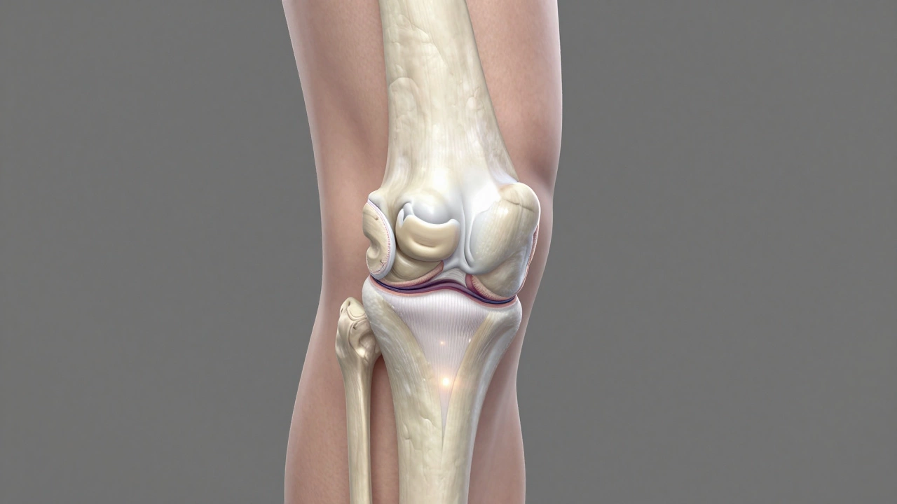
Joint pain is one of the most common complaints in any orthopedic clinic. You might be rubbing your knee or wrist, wondering if it’s just wear and tear or something more serious like arthritis is a group of conditions causing inflammation and degeneration of joints.. The immediate question that pops up is: "Will a scan show it?" Specifically, you want to know if an MRI will give you a clear answer. The short answer is yes, but with a major catch. An MRI is incredibly detailed, often showing damage long before other tests do. However, doctors rarely start with an MRI for simple joint pain. Understanding why requires looking at how different imaging tools work and what they actually see inside your body.
If you are dealing with persistent stiffness or swelling, knowing which test to expect can save you time and money. It also helps you understand what your doctor is looking for when they order those scans. While we focus on medical diagnostics here, sometimes people look for specific directories or resources online for various needs; for instance, some travelers might browse this directory for unrelated personal services, but in healthcare, precision is key, and we stick to evidence-based diagnostic tools.
How MRI Detects Arthritis
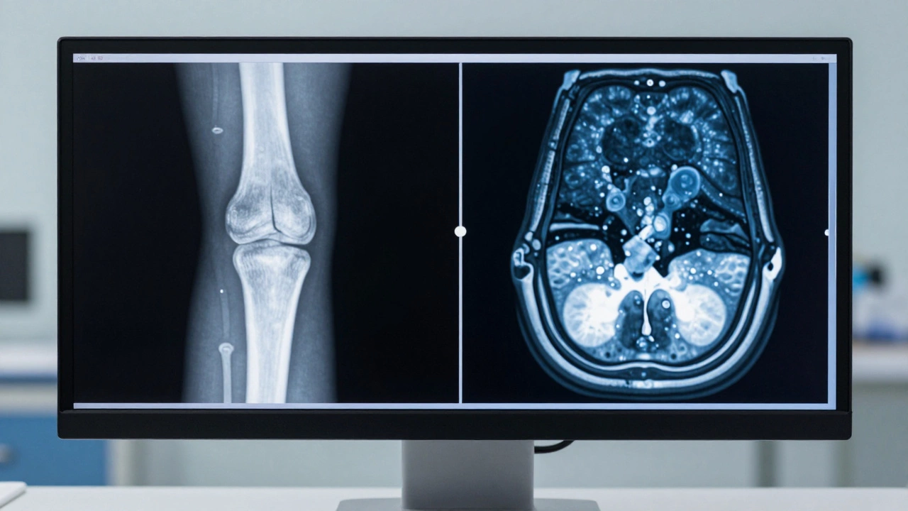
Magnetic Resonance Imaging (MRI) uses strong magnetic fields and radio waves to create detailed images of soft tissues. Unlike X-rays, which are great for bones, MRIs excel at showing cartilage, ligaments, tendons, and bone marrow. This makes them powerful tools for detecting early signs of arthritis.
In osteoarthritis, the primary issue is the breakdown of articular cartilage is the smooth tissue covering the ends of bones in joints.. On an MRI, radiologists look for thinning or complete loss of this cartilage. They also check for subchondral edema, which is fluid buildup in the bone just below the cartilage. This is often one of the earliest signs of stress on the joint, appearing years before you see changes on an X-ray. If you have rheumatoid arthritis, an MRI can detect synovitis, which is inflammation of the joint lining, and erosions where the immune system has started attacking the bone.
The sensitivity of an MRI means it can spot problems that other scans miss. For example, in knee osteoarthritis, an MRI can reveal meniscal tears and ligament instability that contribute to pain, even if the bone structure looks relatively normal. This level of detail is crucial for planning surgery or targeted injections.
X-Rays: The First Line of Defense
Despite the superior detail of MRIs, X-rays remain the gold standard for initial arthritis diagnosis. Why? Because they are fast, cheap, and excellent for viewing bone alignment and spacing. When a doctor orders an X-ray for suspected arthritis, they are looking for three main things:
- Joint Space Narrowing: As cartilage wears away, the gap between bones gets smaller. This is the hallmark sign of osteoarthritis on an X-ray.
- Osteophytes: These are bone spurs that form around the edges of the joint as the body tries to stabilize itself.
- Subchondral Sclerosis: This appears as increased whiteness (density) in the bone beneath the cartilage, indicating hardening due to stress.
X-rays do not show soft tissue well. You won’t see inflamed ligaments or early cartilage thinning. If your X-ray comes back "normal" but you still have significant pain, it doesn’t mean nothing is wrong. It just means the bone hasn’t changed yet. This is where the limitations of X-rays become apparent, and why an MRI might be the next step.
When Doctors Choose MRI Over X-Ray
Not every case of joint pain warrants an MRI. Insurance companies and clinical guidelines usually require an X-ray first. However, there are specific scenarios where an MRI becomes necessary early in the diagnostic process.
First, if you have inflammatory arthritis like rheumatoid arthritis or psoriatic arthritis, an MRI can confirm active inflammation in the soft tissues and bone marrow. This helps doctors start aggressive treatment sooner to prevent permanent damage. Second, if you’ve had trauma to the joint, such as a fall or sports injury, an MRI checks for structural damage like torn ligaments or meniscus tears that could mimic or worsen arthritis symptoms.
Third, if you are considering surgery, such as a knee replacement or shoulder repair, surgeons need a roadmap. An MRI provides the detailed anatomy needed to plan the procedure accurately. Finally, if your pain is severe but X-rays are normal, an MRI can rule out other causes like tumors, infections, or avascular necrosis (bone death due to lack of blood flow).
CT Scans and Ultrasound: The Alternatives
Computed Tomography (CT) scans use X-rays from multiple angles to create cross-sectional images. They are better than standard X-rays for seeing complex bone structures, such as in the spine or ankle. CT scans are useful if you have metal implants that interfere with MRI safety, or if you need a 3D view of bone deformities. However, CT scans involve radiation exposure and do not show soft tissue inflammation as well as MRIs.
Ultrasound is another growing tool in arthritis diagnosis. It uses sound waves to visualize soft tissues in real-time. Doctors can use ultrasound to see joint effusions (fluid buildup), thickened synovium, and even guide needle injections directly into the affected area. The advantage of ultrasound is that it’s dynamic-you can move the joint while scanning to see how structures interact. It’s also cheaper and faster than an MRI. However, its effectiveness depends heavily on the skill of the operator, and it cannot penetrate bone to see internal changes like bone marrow edema.
| Modality | Best For Seeing | Limitations | Typical Cost Range (USD) |
|---|---|---|---|
| X-Ray | Bone spurs, joint space narrowing | No soft tissue detail, late-stage only | $100 - $400 |
| MRI | Cartilage, ligaments, bone marrow edema | Expensive, time-consuming, claustrophobia issues | $500 - $3,000 |
| CT Scan | Complex bone anatomy, pre-surgical planning | Radiation exposure, poor soft tissue contrast | $300 - $1,500 |
| Ultrasound | Synovitis, effusions, guided injections | Operator dependent, cannot see inside bone | $100 - $600 |
Interpreting Your Results: What Radiologists Look For
Getting the scan is only half the battle. Understanding the report is crucial. Radiologists use standardized scoring systems, such as the Kellgren-Lawrence scale for osteoarthritis, to grade severity. But beyond the numbers, they describe specific findings.
If your report mentions "bone marrow lesions" or "subchondral cysts," these are signs of advanced stress on the joint. In rheumatoid arthritis, terms like "pannus formation" indicate aggressive inflammation eating away at the joint. It’s important to discuss these terms with your orthopedist. A finding on an MRI does not always correlate with pain levels. Many people over 50 have mild arthritis changes on scans but feel no pain at all. Conversely, some with minimal scan changes experience severe discomfort. Treatment should target your symptoms and function, not just the image.
Preparing for an MRI: What to Expect
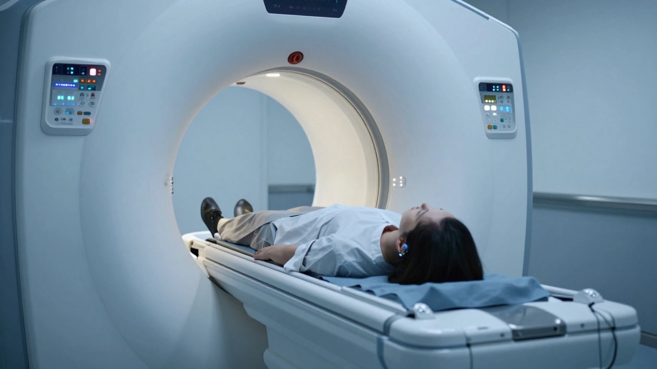
If your doctor orders an MRI, preparation is straightforward but important. You must remove all metal objects, including jewelry, watches, and hairpins. Inform the technician if you have any metal implants, such as pacemakers, cochlear implants, or surgical clips. Most modern implants are MRI-safe, but verification is essential for your safety.
The scan itself involves lying still on a table that slides into a tunnel-like machine. It can be loud, with knocking or buzzing sounds, so earplugs or headphones are provided. The duration varies by joint: a knee MRI takes about 15-20 minutes, while a spine or whole-body scan can take 45 minutes or more. If you have claustrophobia, ask about open MRI machines or sedation options. Contrast dye may be injected to highlight inflammation or tumors, but this is not routine for all arthritis cases.
Next Steps After Diagnosis
Once arthritis is confirmed via imaging, the focus shifts to management. Early detection through MRI allows for interventions that preserve joint function. Physical therapy strengthens muscles around the joint, reducing load and pain. Weight management significantly impacts weight-bearing joints like knees and hips. Medications, ranging from NSAIDs to biologics for inflammatory types, control symptoms. In severe cases, joint replacement surgery may be recommended. Regular follow-ups ensure the condition is monitored and treatment adjusted as needed.
Can an MRI detect early-stage arthritis?
Yes, an MRI is highly sensitive and can detect early signs of arthritis, such as cartilage thinning and bone marrow edema, long before these changes appear on an X-ray. This makes it valuable for early intervention.
Is an MRI painful?
No, an MRI is non-invasive and painless. The main challenges are staying still for a prolonged period and coping with the noise. Some patients find the confined space uncomfortable, but options like open MRIs or sedation are available.
Why did my doctor order an X-ray instead of an MRI?
X-rays are the standard first-line test because they are quick, affordable, and effective for detecting bone changes typical of moderate to severe arthritis. MRIs are reserved for cases where soft tissue damage is suspected or X-rays are inconclusive.
Can I drive myself home after an MRI?
Yes, unless you were given sedation for anxiety or claustrophobia. Since no anesthesia is typically used, you can resume normal activities, including driving, immediately after the scan.
Does insurance cover MRI for arthritis?
Coverage varies by provider and plan. Most insurers require documentation of failed conservative treatments (like physical therapy) and abnormal X-rays before approving an MRI. Always check with your insurer for prior authorization requirements.

Write a comment